


Collagen Loss: Causes, Signs, and How to Slow It
Collagen loss is the progressive reduction and fragmentation of the skin’s main structural protein network, leading to wrinkles, sagging, and volume loss. It starts in your mid-20s and accelerates with UV exposure, smoking, excess sugar, and the enzyme activity of matrix metalloproteinases (MMPs). Two actions you can take today: apply a broad-spectrum SPF 30+ sunscreen every morning and stop smoking. Both have the strongest evidence of any single intervention for slowing collagen degradation.
- Intrinsic aging: Collagen production declines naturally from early adulthood onward, driven by reduced fibroblast activity and hormonal shifts.
- Extrinsic damage: UV radiation, cigarette smoke, glycation from sugar, alcohol, and pollution all accelerate breakdown.
- Enzyme activity: MMPs (MMP-1, MMP-8, MMP-13) actively cleave collagen fibers, and their activity rises sharply with UV exposure and inflammation.
Pro Tip: Apply a vitamin C serum before your SPF every morning. Vitamin C neutralizes free radicals that trigger MMP activity, and sunscreen blocks the UV that activates those enzymes in the first place. Together, they address two of the three main degradation drivers simultaneously.
Key Takeaways
Collagen loss is an active, enzyme-driven process that begins in early adulthood and accelerates with UV exposure, smoking, excess sugar, and hormonal changes, but consistent prevention and evidence-backed topicals can meaningfully slow its progression.
| Point | Details |
|---|---|
| Collagen loss starts early | Production begins declining in the mid-20s; menopause and andropause accelerate the rate further. |
| UV and smoking are top drivers | Both strongly upregulate MMP enzymes that actively cleave collagen fibers; daily SPF 30+ and smoking cessation have the strongest prevention evidence. |
| Retinoids are the best-evidenced topical | Retinoids increase collagen gene transcription and reduce MMP expression; expect 12 weeks of consistent use before visible improvement. |
| No blood test measures skin collagen | Clinicians use visual grading, elasticity devices, ultrasound, and biopsy; commercial “collagen level” tests are not validated. |
| Cellure’s Complete Skin Repair Kit | Combines peptides, tranexamic acid, and hydrating actives in a science-backed routine targeting collagen remodeling and skin renewal. |
Table of Contents
- What collagen is and why your skin depends on it
- How collagen is actually broken down at the molecular level
- What causes collagen loss to speed up?
- How collagen loss shows up on your skin and body
- How clinicians assess collagen loss (and why there is no blood test for it)
- Everyday habits that protect your collagen
- Which topical ingredients actually support collagen?
- In-office procedures for collagen stimulation and volume replacement
- What the evidence actually says about collagen supplements
- When collagen loss signals something more serious
- How collagen loss affects mobility and quality of life over time
- Do collagen loss patterns differ by skin type and ethnicity?
- What realistic collagen repair actually looks like
- Cellure’s Complete Skin Repair Kit fits an evidence-based collagen-support plan
- Sources
What collagen is and why your skin depends on it
Collagen makes up roughly 30% of the body’s total protein, and in the skin it functions as the structural scaffold that gives tissue its tensile strength, volume, and resilience. Without adequate collagen, skin loses its ability to snap back after stretching and begins to sag.
The skin has two main layers relevant here. The epidermis is the thin outer surface you can see and touch. The dermis sits beneath it and is where collagen actually lives, woven into a dense fibrous matrix alongside elastin and hyaluronic acid. This distinction matters for topical treatments: most serums and creams cannot penetrate deep enough to directly rebuild dermal collagen. What they can do is signal fibroblasts (the cells that produce collagen) or protect existing fibers from damage.
Key collagen types in skin:
- Type I: The dominant structural collagen, accounting for most of the dermis’s dry weight; responsible for tensile strength and firmness.
- Type III: More flexible and abundant in younger skin; its ratio to Type I shifts with age, contributing to reduced elasticity.
Beyond skin, collagen is the primary structural protein in cartilage, bone, and tendons. That systemic role explains why collagen decline eventually shows up as joint stiffness and reduced bone density, not just wrinkles.
How collagen is actually broken down at the molecular level
Collagen loss is not simply a slowdown in production. It is an active, enzyme-driven demolition process running in parallel with declining synthesis.

The main enzymes responsible are the matrix metalloproteinases (MMP-1, MMP-8, and MMP-13), a family of collagenases that cleave the triple-helix structure of fibrillar collagen. UV radiation is one of the strongest known triggers for MMP upregulation. A single unprotected sun exposure can measurably increase MMP activity in dermal tissue within hours. Chronic UV exposure compounds this into years of cumulative fiber destruction.
On the production side, dermal fibroblasts lose both their numbers and their synthetic capacity with age. Biopsy studies confirm that elderly skin contains fewer fibroblasts than younger skin, and those that remain produce less collagen per cell. There is also a mechanical feedback loop at work: fragmented, disorganized collagen provides a weaker physical scaffold, which in turn reduces the mechanical stimulation fibroblasts need to stay active. Less stimulation means less collagen output, which means more fragmentation. The cycle accelerates itself.
Two additional accelerants deserve attention:
- Inflammaging: Low-grade, chronic inflammation that accumulates with age elevates cytokine levels that upregulate MMP expression and suppress collagen gene transcription. Poor sleep, obesity, and chronic stress all feed this state.
- Glycation and AGEs: Excess dietary sugar binds to collagen fibers through a process called glycation, forming advanced glycation end-products (AGEs) that stiffen and disorganize the collagen matrix, making it more susceptible to enzymatic cleavage. High-sugar diets directly compromise long-term skin resilience through this mechanism.
Research into peptide and peptidomimetic inhibitors of MMP activity is an active area in cosmeceutical science, with the goal of preserving fibrillar collagen by blocking the enzymes that degrade it.
What causes collagen loss to speed up?
Collagen decline has two categories of drivers: intrinsic factors you cannot change and extrinsic factors you largely can.
Intrinsic causes:
- Chronological aging (production decline begins in the mid-20s)
- Genetic predisposition to faster fibroblast senescence
- Hormonal changes: estrogen supports collagen synthesis, so women experience an accelerated decline around menopause, with some estimates suggesting a significant drop in dermal collagen density in the first years after menopause
- Andropause in men produces a slower but real decline in testosterone-related collagen support
Extrinsic causes (modifiable):
- UV radiation: The single largest extrinsic driver; activates MMPs and generates reactive oxygen species that damage collagen directly
- Smoking: Reduces blood flow to the dermis, elevates MMP activity, and depletes vitamin C needed for collagen synthesis
- Excess sugar: Drives glycation and AGE formation, stiffening and fragmenting collagen fibers
- Alcohol: Dehydrates tissue and impairs the liver’s ability to process collagen precursors
- Air pollution: Particulate matter and ozone generate oxidative stress in the dermis
- Poor sleep: Elevates cortisol and inflammatory markers that suppress collagen synthesis
- Chronic inflammation: Sustained MMP upregulation from any inflammatory source degrades collagen faster than it can be replaced
UV and smoking are the highest-priority targets. Both have strong mechanistic and epidemiological evidence linking them to accelerated collagen degradation, and both are fully modifiable. Lifestyle factors including smoking, excess alcohol, and high-sugar diets accelerate collagen fragmentation and fibroblast impairment, compounding the natural rate of intrinsic decline.
Pro Tip: Address risk factors in order of impact. Sunscreen every day comes first. Quitting smoking is second. Reducing added sugar is third. Targeted topical actives come after these foundations are in place — not instead of them.
How collagen loss shows up on your skin and body
The signs of collagen depletion fall into two categories: surface textural changes and structural changes. They look different and mean different things.
Surface textural changes (early signs):
- Fine lines, especially around the eyes and mouth
- Crepey texture on the neck, décolletage, and inner arms
- Reduced skin “bounce” when pressed
Structural changes (later or more significant loss):
- Deep wrinkles and folds (nasolabial folds, marionette lines)
- Sagging along the jawline and cheeks
- Hollowing under the eyes and in the temples (volume loss, not just surface wrinkling)
- Increased skin fragility and easy bruising
- Slower wound healing and thinner skin overall
The distinction between surface texture and structural volume matters because they respond to different interventions. Fine lines can improve with topical retinoids and hydration. Volume loss from structural collagen depletion generally requires either procedural stimulation (microneedling, radiofrequency) or replacement (dermal fillers).
Beyond skin, collagen loss in connective tissues shows up as joint stiffness, reduced cartilage cushioning, and in severe or prolonged cases, increased fracture risk from reduced bone collagen matrix. These non-skin signs are worth noting because they confirm that collagen decline is a systemic process, not a cosmetic inconvenience.

How clinicians assess collagen loss (and why there is no blood test for it)
There is no routine blood test that measures your skin’s collagen level. Serum markers like procollagen peptides exist in research settings, but they reflect whole-body collagen turnover, not the specific density or quality of collagen in your dermis. Clinicians rely on observable and measurable clinical indicators instead.
Common assessment approaches:
- Visual and photographic grading: Standardized scales (such as the Glogau scale for photoaging) allow clinicians to document and track changes over time.
- Cutometer or skin elasticity devices: Measure how much skin deforms under suction and how quickly it returns to baseline, giving a functional proxy for collagen and elastin integrity.
- High-frequency ultrasound: Can image dermal thickness and density non-invasively; used in research and some clinical settings to quantify structural changes.
- Skin biopsy and histology: The gold standard for measuring collagen fiber density and organization; reserved for suspected connective tissue disease or unusual clinical presentations.
Be cautious about commercial “collagen level tests” marketed directly to consumers. These are not validated clinical tools, and their results are not comparable to dermatological assessment.
When to seek objective clinical assessment:
- Rapid or unexplained skin thinning over weeks to months
- Wound-healing problems (cuts or bruises that take unusually long to resolve)
- Suspected connective tissue disease (Ehlers-Danlos syndrome, lupus, scleroderma)
- Significant changes in joint mobility alongside skin changes
Everyday habits that protect your collagen
Prevention has the strongest evidence base of any collagen-support strategy. The following measures are ranked roughly by evidence strength.
- Daily broad-spectrum sunscreen (SPF 30 or higher) — strong evidence. Apply enough to cover all exposed skin (about a nickel-sized amount for the face alone) and reapply every two hours outdoors. Harvard’s Nutrition Source lists sunscreen as one of the most consistent behavioral defenses against collagen degradation.
- Stop smoking — strong evidence. Smoking impairs dermal circulation, elevates MMPs, and depletes the vitamin C required for collagen synthesis. No topical product compensates for active smoking.
- Limit added sugar — moderate evidence. Reducing dietary sugar slows AGE formation and glycation-related collagen stiffening.
- Adequate protein intake — moderate evidence. Collagen synthesis requires amino acids, particularly glycine, proline, and hydroxyproline. Aim for adequate total protein from whole food sources.
- Vitamin C from food or supplements — moderate evidence. Vitamin C is a required cofactor for the enzymes that stabilize collagen’s triple-helix structure. Citrus, bell peppers, and leafy greens are reliable sources.
- Adequate sleep each night — moderate evidence. Sleep is when growth hormone peaks and tissue repair occurs; chronic short sleep elevates cortisol and inflammatory markers that suppress collagen synthesis.
- Regular exercise — moderate evidence. Resistance training and aerobic exercise support circulation and reduce systemic inflammation, both of which benefit fibroblast function.
- Limit excess alcohol — moderate evidence. Heavy alcohol use dehydrates tissue and impairs collagen precursor processing.
- Manage chronic stress — moderate evidence. Sustained cortisol elevation suppresses collagen gene expression.
A Mediterranean-style dietary pattern (high in vegetables, legumes, fish, and olive oil; low in ultra-processed foods and added sugar) aligns well with most of these nutritional recommendations and has observational support for skin health outcomes.
Pro Tip: For sunscreen, a mineral formula with zinc oxide works well for sensitive skin and doubles as a physical barrier against pollution. Apply it as the last step in your morning routine, after any vitamin C serum, to lock in both layers of protection.
Which topical ingredients actually support collagen?
Several topical actives have clinical evidence for stimulating collagen remodeling or protecting existing fibers. Evidence levels vary considerably.
- Retinoids (retinol, tretinoin) — strong evidence. Retinoids increase collagen gene transcription in fibroblasts and reduce MMP expression. Prescription tretinoin has the most robust data. Over-the-counter retinol is effective at higher concentrations (0.3–1%) but takes longer. Start low (0.025–0.05% tretinoin or 0.1% retinol), use at night, and always follow with SPF the next morning. Irritation is common initially; introduce slowly.
- Topical vitamin C (L-ascorbic acid) — moderate to strong evidence. Stabilizes collagen synthesis as a cofactor and neutralizes free radicals that trigger MMP activity. Effective concentrations are 10–20%. Formulation stability matters: look for products in opaque, airtight packaging.
- Peptides — emerging to moderate evidence. Signal peptides (like Matrixyl/palmitoyl pentapeptide-4) and carrier peptides (like copper peptides/GHK-Cu) have shown fibroblast-stimulating activity in cell studies and some clinical trials. The role of peptides in skin repair is an active area of cosmeceutical research, with science-backed peptide strategies showing meaningful results for collagen remodeling.
- Niacinamide — moderate evidence. Reduces inflammation, supports the skin barrier, and has some evidence for stimulating collagen synthesis at concentrations of 5% or higher.
- Topical antioxidants (vitamin E, ferulic acid, resveratrol) — moderate evidence for protection. Primarily work by neutralizing oxidative stress rather than directly stimulating collagen production; most effective when combined with vitamin C and sunscreen.
Realistic expectations: topical actives stimulate remodeling over months, not days. They can meaningfully improve fine lines and skin texture, but they do not replace large-volume structural collagen loss. For that, procedural options are more appropriate.
In-office procedures for collagen stimulation and volume replacement
Procedural options split into two strategies: stimulating your skin to produce new collagen, or replacing lost volume directly.
Stimulation-based approaches:
- Microneedling: Creates controlled micro-injuries that trigger a wound-healing response and fibroblast activation. Multiple sessions (typically 3–6) are needed; downtime is 24–72 hours of redness. Evidence for collagen induction is solid.
- Radiofrequency (RF): Delivers heat energy to the dermis, denaturing existing collagen and stimulating new fiber formation. Non-ablative RF has minimal downtime; ablative fractional RF has more but stronger results.
- Laser resurfacing: Ablative lasers (CO2, Er:YAG) remove the epidermis and heat the dermis, producing significant collagen remodeling. Downtime ranges from 5–14 days. Non-ablative lasers have less downtime but more modest results.
- Platelet-rich plasma (PRP): Uses growth factors from your own blood to stimulate fibroblast activity. Evidence is promising but more variable than RF or laser; often combined with microneedling.
Volume replacement:
- Dermal fillers (hyaluronic acid, calcium hydroxylapatite, poly-L-lactic acid): Do not stimulate collagen directly (though some biostimulatory fillers do over time) but restore volume immediately. Results last 6–24 months depending on the product.
| Approach | Mechanism | Sessions needed | Downtime | Best for |
|---|---|---|---|---|
| Microneedling | Wound-healing stimulation | 3–6 | 24–72 hours | Texture, fine lines |
| Radiofrequency | Thermal collagen remodeling | 1–4 | Minimal to moderate | Laxity, firmness |
| Ablative laser | Resurfacing + dermal heating | 1–2 | 5–14 days | Deep lines, texture |
| PRP | Growth factor stimulation | 3–4 | Minimal | Tone, early laxity |
| Dermal fillers | Volume replacement | 1 | Minimal | Volume loss, deep folds |
Always verify that your provider is a licensed dermatologist or plastic surgeon with documented experience in the specific device or technique. Clinical evidence for these procedures varies by modality, and outcomes depend heavily on operator skill.
What the evidence actually says about collagen supplements
Oral collagen peptides (hydrolyzed collagen) are among the most widely sold supplements in the U.S. The evidence is real but modest and worth understanding clearly.
- Skin elasticity and hydration: Randomized controlled trials have reported modest improvements in skin elasticity and hydration with oral collagen peptides, but many trials are small, short in duration, and some are industry-funded. Independent replication is still limited.
- Joint mobility: Some trials in athletes and older adults report reduced joint discomfort with collagen peptide supplementation, but the evidence base is smaller and less consistent than for skin outcomes.
- Typical doses in trials: Studies generally use 2.5–10 grams of hydrolyzed collagen peptides per day, often for 8–12 weeks. Vitamin C is frequently co-administered because it is required for collagen synthesis.
- Safety: Collagen supplements are generally well-tolerated. Potential concerns include allergic reactions in people sensitive to the source animal (bovine, marine, porcine), and those who are pregnant or have kidney disease should consult a clinician before use.
The honest summary: supplements are unlikely to cause harm at typical doses and may offer modest skin and joint benefits for some people. They are not a substitute for sunscreen, a retinoid, or smoking cessation, all of which have far stronger evidence. More independent, long-term trials are needed before strong recommendations can be made.
When collagen loss signals something more serious
Most collagen loss is a normal part of aging, manageable with lifestyle and topical strategies. Some presentations warrant clinical attention.
Red flags that warrant prompt evaluation:
- Rapid, unexplained skin thinning over weeks to months
- Easy bruising without injury, or bruises that spread unusually
- Wounds that fail to close or heal within a normal timeframe
- Severe joint hypermobility, pain, or instability alongside skin changes
- Skin that tears easily with minor trauma
- Suspected autoimmune disease (joint swelling, rash, fatigue, dry eyes/mouth)
These signs can indicate connective tissue disorders (Ehlers-Danlos syndromes, Marfan syndrome), autoimmune conditions (lupus, scleroderma), or nutritional deficiencies severe enough to impair collagen synthesis (scurvy from vitamin C deficiency, though rare in the U.S.).
What to expect at a clinical visit: A dermatologist or rheumatologist will take a detailed history, examine the skin and joints, and may order imaging or a skin biopsy if a connective tissue disease is suspected. Bring dated photographs documenting the changes, note when symptoms began, and write down any medications or supplements you are taking. Asking specifically about collagen-related conditions helps the clinician focus the evaluation.
This article provides general health information and is not a substitute for professional medical advice. Consult a qualified clinician for diagnosis or treatment of any medical condition.
How collagen loss affects mobility and quality of life over time
The long-term consequences of collagen decline extend well beyond cosmetic concerns. Collagen is the primary structural protein in cartilage, the tissue that cushions joints. As cartilage collagen degrades with age, joint surfaces become less protected, contributing to the stiffness and discomfort associated with osteoarthritis. Tendons and ligaments, also collagen-dense, lose tensile strength and become more injury-prone.
Bone matrix is roughly one-third collagen by weight. When collagen quality deteriorates, bone becomes more brittle even when mineral density appears adequate on a standard scan. This partly explains why fracture risk increases with age independently of bone density measurements.
For mobility and daily function, the cumulative picture is significant. Reduced joint cushioning limits range of motion. Weaker tendons increase injury risk during physical activity. Thinner, more fragile skin heals more slowly after minor injuries, which matters especially for older adults. Together, these changes can meaningfully reduce independence and quality of life, which is why collagen support strategies (exercise, protein intake, vitamin C, and in some cases supplementation) are worth taking seriously as a long-term investment, not just a cosmetic one.
Do collagen loss patterns differ by skin type and ethnicity?
Yes, and the differences are clinically meaningful. Skin with higher melanin content (Fitzpatrick types IV–VI) generally has a thicker dermis and denser collagen network at baseline, which provides some structural advantage against early photoaging. People with darker skin tones often develop fine lines and surface wrinkles later than those with lighter skin, but they are not immune to collagen loss. They tend to experience it differently: volume loss and skin laxity can become apparent before surface wrinkling does, which means the signs are sometimes missed or attributed to other causes.
Lighter skin (Fitzpatrick types I–III) is more susceptible to UV-induced MMP activation and photoaging, meaning extrinsic collagen degradation tends to manifest earlier and more visibly as fine lines and texture changes.
Hormonal factors affect all skin types, but the rate of post-menopausal collagen decline appears consistent across ethnicities. What differs is the starting point and the dominant clinical presentation. For practical purposes, this means prevention strategies (sunscreen, retinoids, lifestyle) are universally relevant, but the signs to watch for and the timeline of visible change vary by individual skin biology.
What realistic collagen repair actually looks like
The gap between what the skincare industry promises and what the science actually supports is wide. Most products imply rapid, dramatic restoration. The biology tells a different story.
Collagen remodeling is slow. Retinoids, the best-evidenced topical actives, typically require 12 weeks of consistent use before measurable changes in collagen density appear in biopsy studies. Microneedling results continue to develop for 3–6 months after a session. Even the most aggressive in-office procedures work by triggering a wound-healing cascade that unfolds over months, not days.
What this means practically: the people who see real results are the ones who build consistent routines and stick with them. A daily SPF, a retinoid three nights a week, and adequate protein and sleep will outperform any single “miracle” product used sporadically. Procedures add meaningful benefit on top of that foundation, but they are not a shortcut around it.
The other underappreciated point is that collagen quality matters as much as quantity. Aged collagen becomes fragmented and disorganized, reducing elasticity even when some collagen remains. Interventions that improve collagen organization (retinoids, RF, microneedling) can restore function even without dramatically increasing total collagen volume. That is a more realistic and achievable goal than “replacing” lost collagen wholesale.
Prioritize prevention first, then evidence-backed topicals, then procedures if needed. Expect months, not weeks. And be skeptical of any product that claims to “rebuild” collagen overnight.
Cellure’s Complete Skin Repair Kit fits an evidence-based collagen-support plan
Sunscreen, retinoids, and lifestyle changes form the foundation. The next layer is a targeted topical routine built around actives that have real mechanistic support for collagen remodeling. That is where Cellure’s Complete Skin Repair Kit fits in.
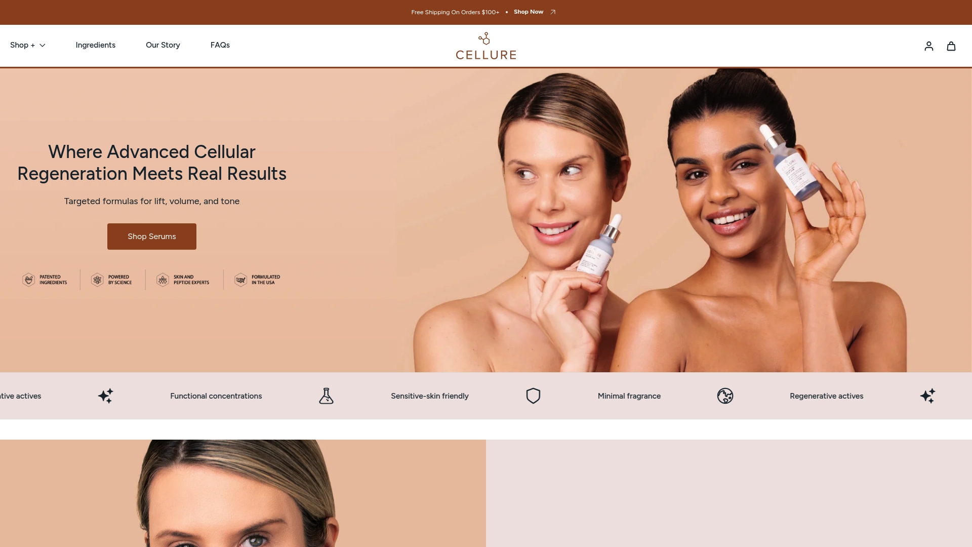
The kit combines peptides, tranexamic acid, hyaluronic acid, and hydrating repair actives in formulations designed for cellular-level skin renewal. Peptides signal fibroblasts to support collagen production; tranexamic acid addresses uneven tone that often accompanies structural aging; hyaluronic acid restores surface hydration that collagen loss depletes. The formulas are designed to be gentle enough for sensitive skin while delivering the concentrations that clinical evidence supports.
Results vary by individual. For medical concerns or significant collagen-related symptoms, consult a dermatologist. Explore the Complete Skin Repair Kit to see the full ingredient list and how each active fits into a science-backed routine.
Sources
The following sources were used in this article and are worth reading directly for deeper mechanistic detail or clinical guidance.
- Collagen degradation pathways review — PMC
- Collagen: What It Is, Types, Function & Benefits — Cleveland Clinic
- Collagen • The Nutrition Source — Harvard T.H. Chan School of Public Health
- Collagen: Benefits, Side Effects, and Caution — Healthline
Recommended
Share