


Hyperpigmentation: Causes, Diagnosis, and Safe Treatment
Hyperpigmentation is a skin condition in which excess or clumped melanin creates patches or spots darker than the surrounding skin. The two most important steps you can take right now: apply a broad-spectrum SPF 30+ sunscreen every morning, and see a dermatologist if any discoloration is new, rapidly changing, or widespread.
Immediate actions:
- Start daily broad-spectrum sunscreen (SPF 30 or higher), reapplied every two hours outdoors
- Avoid picking at acne, scabs, or irritated skin to reduce post-inflammatory darkening
- Note any recent medication changes, hormonal shifts, or skin trauma that may be driving pigment changes
- Book a dermatologist appointment for lesions with irregular borders, rapid change, or unexplained diffuse darkening
Pro Tip: Apply sunscreen as the very last step in your morning routine, after moisturizer and before makeup, and reapply at midday even on cloudy days. UV radiation passes through clouds and glass.
Key Takeaways
Hyperpigmentation is a chronic, often relapsing condition where sun protection is the single most important daily habit, and treatment works best when matched to the specific type and pigment depth.
| Point | Details |
|---|---|
| Core definition | Excess or clumped melanin causes darker patches; usually painless but can signal systemic disease. |
| Primary cause | UV/sun exposure is the most common trigger; hormones, inflammation, and medications also drive pigment changes. |
| First-line prevention | Daily broad-spectrum SPF 30+ sunscreen is the single most effective step, supporting every other treatment. |
| Safe treatment sequence | Diagnose the type first, then add photoprotection, topical agents, and professional procedures only as needed. |
| Cellure’s role | The Complete Skin Repair Kit pairs tranexamic acid with peptides and polynucleotides for maintenance-phase pigment control and barrier repair. |
Table of Contents
- What is hyperpigmentation, and how does melanin cause it?
- Common types of hyperpigmentation and how to tell them apart
- Why hyperpigmentation happens: UV, hormones, inflammation, and more
- How a dermatologist evaluates and diagnoses hyperpigmentation
- Safe, evidence-based treatment options for hyperpigmentation
- What clinical evidence and 2026 guidelines say about treatment
- How to prevent hyperpigmentation and protect treatment gains
- What to realistically expect from treatment
- When you should see a dermatologist about skin darkening
- A note on what actually matters in long-term pigment management
- Cellure’s approach to maintenance-focused brightening
- Sources
What is hyperpigmentation, and how does melanin cause it?
Hyperpigmentation happens when melanocytes, the pigment-producing cells in the skin’s basal layer, overproduce or unevenly distribute melanin. UV radiation, inflammation, and hormonal signals all trigger melanocytes to ramp up activity, and the resulting pigment accumulates in localized clusters rather than spreading evenly.
Two main melanin types determine visible color: eumelanin (brown to black) and pheomelanin (yellow to red). The depth at which pigment sits matters as much as the amount. Epidermal pigment tends to look brown and responds well to topical agents. Dermal pigment sits deeper, often appears gray or blue-gray, and resists most topical treatments.
- Melanocytes transfer melanin to surrounding keratinocytes via structures called melanosomes
- UV exposure, hormones, and inflammatory mediators (especially prostaglandins and cytokines) all upregulate melanin synthesis
- Visible color ranges from light brown to near-black depending on melanin type, concentration, and skin depth
- Hyperpigmentation is almost always painless and cosmetic, but diffuse or sudden-onset darkening can occasionally signal systemic disease
Common types of hyperpigmentation and how to tell them apart
Identifying the specific type matters because treatment response differs significantly by type and pigment depth. The four types you are most likely to encounter are melasma, post-inflammatory hyperpigmentation (PIH), solar lentigines, and acanthosis nigricans.
Melasma appears as symmetrical, blotchy brown or gray-brown patches, typically on the cheeks, forehead, upper lip, and chin. Hormonal changes, including pregnancy and oral contraceptives, are the dominant triggers, and UV exposure reliably worsens it. Melasma is often mixed epidermal and dermal, which makes it one of the more stubborn types to treat.
Post-inflammatory hyperpigmentation follows any skin injury or inflammation: acne, burns, eczema, or even an aggressive facial treatment. PIH commonly persists unless both inflammation and barrier damage are addressed, and darker skin tones are disproportionately affected.
Solar lentigines (age spots or sunspots) are small, well-defined brown spots that develop on sun-exposed areas, particularly the face, hands, and shoulders. They are purely epidermal and generally respond well to topical brighteners and light-based procedures.

Acanthosis nigricans is a distinct pattern: velvety, dark thickening in skin folds such as the neck, armpits, and groin. Unlike the other types, it often signals an underlying metabolic or endocrine condition and warrants medical evaluation rather than cosmetic treatment.
| Type | Typical location | Common triggers | Treatment responsiveness |
|---|---|---|---|
| Melasma | Cheeks, forehead, upper lip | Hormones, UV | Moderate; often recurs |
| PIH | Site of prior injury/acne | Inflammation, trauma | Good if epidermal; slower if dermal |
| Solar lentigines | Face, hands, shoulders | Cumulative UV | Good with topicals and lasers |
| Acanthosis nigricans | Neck, armpits, groin | Metabolic/endocrine | Treat underlying cause first |
Self-assessment is reasonable for stable, well-defined spots with a clear trigger. Seek professional evaluation for anything with irregular borders, rapid change, or no obvious cause.
Why hyperpigmentation happens: UV, hormones, inflammation, and more
Sun exposure is the most common external cause: UV radiation directly stimulates melanocytes to produce more melanin as a protective response, and cumulative exposure over years produces permanent changes in melanocyte behavior.
Beyond UV, the major drivers include:
- Hormonal changes: Estrogen and progesterone sensitize melanocytes, which is why melasma spikes during pregnancy and with hormonal contraceptive use
- Inflammation and injury: Acne, eczema, cuts, chemical burns, and even aggressive skincare procedures can all trigger PIH
- Medications: Certain antibiotics (tetracyclines), antimalarials, chemotherapy agents, and nonsteroidal anti-inflammatory drugs can cause drug-induced pigmentation
- Systemic disease: Addison disease causes diffuse bronze darkening from elevated ACTH; hemochromatosis produces a gray-brown cast; thyroid disorders can also alter pigment distribution
- Genetics: Freckles (ephelides) are largely genetically determined and intensify with UV exposure
- Phytophotodermatitis: Contact with certain plants or fruits (limes, celery, parsnips) followed by sun exposure can cause dramatic, patterned hyperpigmentation that is often mistaken for a bruise or chemical burn
A practical self-check: review any medications started in the past six months, note any recent hormonal changes, and map whether darkened areas correspond to sites of prior acne or skin trauma. Diffuse or unexplained darkening that doesn’t fit these patterns warrants systemic evaluation for conditions like Addison disease or hemochromatosis.
How a dermatologist evaluates and diagnoses hyperpigmentation
Diagnosis is usually clinical, meaning a trained eye and a good history are enough in most cases. Tests are used selectively to determine pigment depth or rule out systemic causes, as the Merck Manual’s professional guidance makes clear.
A clinician will typically ask about:
- Onset and progression (sudden vs. gradual, stable vs. expanding)
- Possible triggers: recent sun exposure, medications, pregnancy, contraceptive use
- Personal and family history of skin conditions or systemic disease
- Any associated symptoms (fatigue, weight changes, joint pain) that might suggest a systemic cause
| Diagnostic tool | What it shows | When it’s used |
|---|---|---|
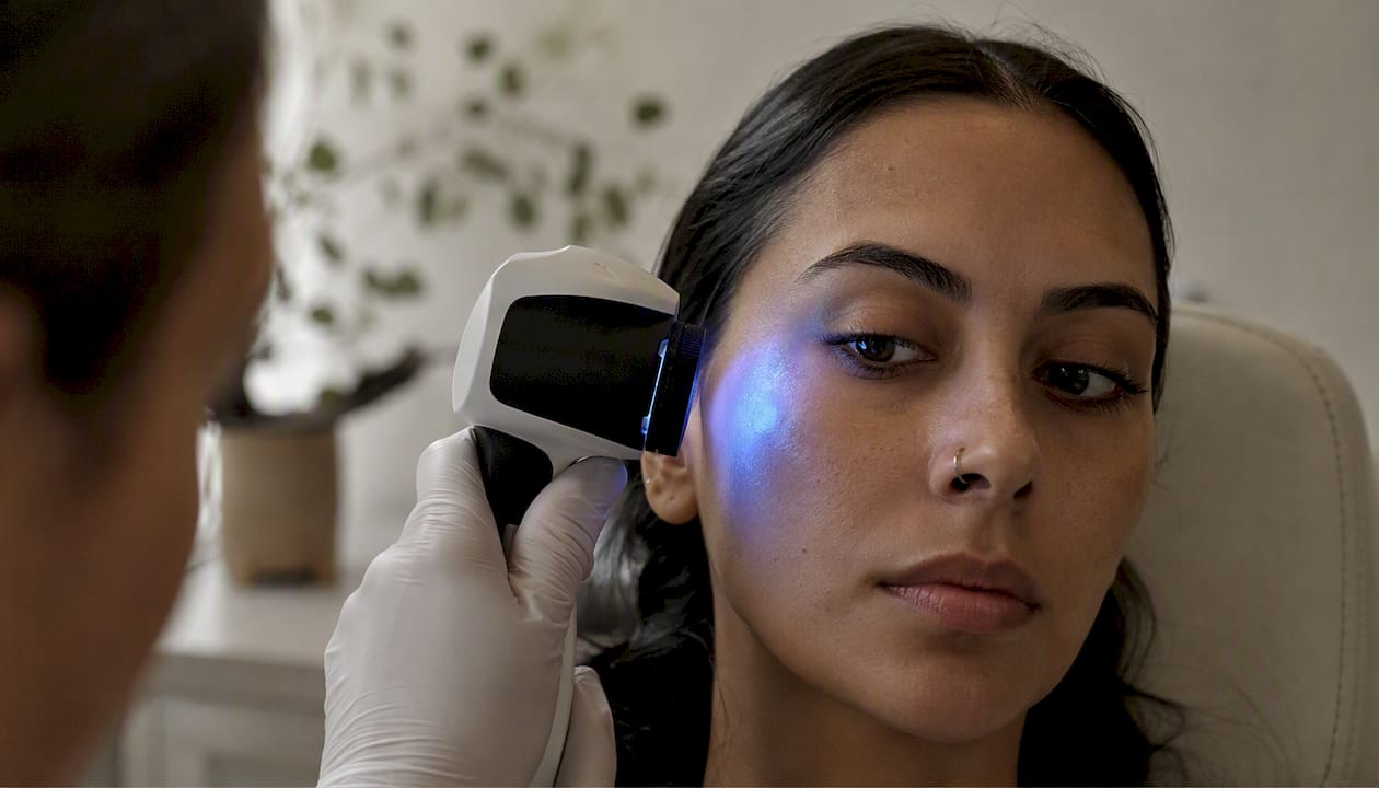
| Wood’s lamp | Epidermal vs. dermal pigment depth | Routine; helps guide treatment choice |
| Dermoscopy | Pigment pattern and distribution | Distinguishing benign from suspicious lesions |
| Skin biopsy | Histological pigment type and depth | Suspected malignancy or atypical presentation |
| Lab testing (CBC, cortisol, thyroid) | Systemic causes of diffuse darkening | Unexplained widespread pigmentation |
Identifying whether pigment is epidermal or dermal via Wood’s lamp substantially changes likely treatment success and expected timeframe. Epidermal pigment fluoresces under Wood’s lamp; dermal pigment does not.
Safe, evidence-based treatment options for hyperpigmentation
The treatment roadmap follows a clear sequence: sun protection first, then topical agents, then professional procedures if needed, then long-term maintenance. Harvard Health and the Cleveland Clinic both emphasize that skipping photoprotection undermines every other intervention.
Topical agents are the first active treatment step:
- Hydroquinone: The most studied depigmenting agent; effective short-term (typically 3–6 months) but not for indefinite use due to risks including exogenous ochronosis with prolonged application
- Azelaic acid: Targets tyrosinase (the enzyme driving melanin synthesis); well tolerated, including during pregnancy
- Retinoids: Accelerate cell turnover and disperse melanin granules; require gradual introduction to avoid irritation-triggered PIH
- Tranexamic acid: Blocks the UV-induced pathway that stimulates melanocytes; increasingly favored as a maintenance agent with a strong safety profile. Cellure’s guide to tranexamic acid for hyperpigmentation covers the mechanism in depth
- Kojic acid and vitamin C: Antioxidant-based brighteners that inhibit melanin oxidation; useful as adjuncts
In-office procedures include chemical peels, laser treatments, intense pulsed light (IPL), and microneedling. These can accelerate results but carry real risks, particularly for darker skin tones.
Dermatologists commonly advise against using aggressive lasers or strong peels as first-line treatment for PIH because procedures that provoke inflammation can worsen pigmentation if not carefully selected. Any ablative or heat-based procedure should be preceded by a period of topical priming and followed by strict photoprotection.
Systemic options (oral tranexamic acid, for example) are used in specific cases under medical supervision. Combination regimens, such as the classic triple combination of hydroquinone, a retinoid, and a topical corticosteroid, are effective short-term but are always intended as a bridge to gentler maintenance therapy.
Pro Tip: Pair any anti-pigment active with a barrier-repair ingredient (ceramides, niacinamide, or peptides) to reduce the irritation that can itself trigger rebound PIH. Anti-inflammatory care and brightening work better together than either does alone.
What clinical evidence and 2026 guidelines say about treatment
Photoprotection is the foundation, and accurate diagnosis guides safe therapy. That is the consistent message across the 2026 Medscape global guideline summary on melanin hyperpigmentation disorders, which recommends individualized treatment plans and explicit caution with ablative interventions due to relapse and worsening risk.
Key guideline takeaways:
- Photoprotection is non-negotiable as both prevention and treatment adjunct
- Treatment selection must match the specific disorder; what works for solar lentigines can worsen melasma
- Hyperpigmentation is often chronic and relapsing; long-term maintenance planning improves outcomes
- Aggressive procedures carry a real risk of worsening pigmentation, particularly in skin of color
- Staged approaches, starting potent agents short-term and transitioning to gentler maintenance actives, reduce adverse events
Effective long-term management addresses the skin microenvironment: calming inflammation, supporting cellular repair, and reinforcing the barrier, rather than only bleaching pigment at the surface. Ingredients like polynucleotides, copper peptides, and growth factors are gaining traction in this maintenance role. The science behind skin brightening explains how these ingredient classes interact in practice.
How to prevent hyperpigmentation and protect treatment gains
Consistent sun protection is the single most effective prevention strategy, full stop. Every other measure builds on it.
- Sunscreen: Broad-spectrum SPF 30 minimum daily; SPF 50 for active outdoor exposure. Reapply every two hours. Tinted mineral sunscreens (with iron oxides) offer additional protection against visible light, which can worsen melasma even without UV
- Physical blockers: Zinc oxide and titanium dioxide sit on the skin’s surface and deflect UV rather than absorbing it; preferred for sensitive skin and post-procedure care
- Avoid picking: Squeezing acne or picking at scabs is one of the most reliable ways to create PIH
- Treat inflammation early: Address eczema flares, rashes, and acne promptly to minimize the inflammatory signal that triggers melanocytes
- Review medications: If a drug is photosensitizing (certain antibiotics, diuretics, retinoids), discuss sun precautions with your prescriber
- Gentle exfoliation: Chemical exfoliants (AHAs, BHAs) at appropriate concentrations support cell turnover without the micro-trauma of physical scrubs
For maintenance, retinoids, tranexamic acid, and antioxidants (vitamin C, niacinamide) are the most evidence-supported choices. Return to your dermatologist if pigmentation returns or worsens despite consistent maintenance.
Pro Tip: Layer actives strategically: apply vitamin C in the morning (antioxidant protection) and retinoids at night (cell turnover). This timing reduces the chance of irritation and maximizes each ingredient’s mechanism.
What to realistically expect from treatment
Many types of hyperpigmentation improve meaningfully with appropriate treatment, but recurrence is common, and gradual is the operative word. Even effective topical regimens typically require several weeks to months for visible lightening, and realistic expectations reduce the temptation to abandon treatment or try unsafe shortcuts.
Epidermal pigment responds faster and more completely than dermal pigment. Early treatment, before pigment migrates deeper, consistently predicts better outcomes. Rigorous photoprotection throughout treatment is not optional; without it, UV exposure restimulates melanocytes and erases progress.
Melasma, in particular, tends to be chronic. Many people manage it rather than cure it, cycling through active treatment phases and maintenance periods. Solar lentigines treated with lasers or cryotherapy often clear well but can recur with continued sun exposure.
When you should see a dermatologist about skin darkening
Some situations call for professional evaluation rather than watchful waiting.
See a dermatologist promptly if you notice:
- A lesion with irregular borders, multiple colors, or asymmetry (rule out melanoma)
- Rapid change in size, shape, or color of any spot
- Widespread or diffuse darkening without a clear trigger (sun, medication, or recent inflammation)
- Darkening accompanied by systemic symptoms: fatigue, unexplained weight loss, joint pain, or abdominal symptoms
- Pigmentation that appears to follow a medication you recently started
- Velvety dark thickening in skin folds (possible acanthosis nigricans, which needs metabolic workup)
When you go, bring a list of all current medications and supplements, and if possible, photos showing the progression of the lesion over time. For anything that looks like it could be melanoma, same-week evaluation is appropriate, not a wait-and-see approach.
A note on what actually matters in long-term pigment management
The skincare industry tends to frame hyperpigmentation as a problem you solve once and move on. Dermatology says otherwise. The Medscape 2026 guideline is explicit: this is often a chronic, relapsing condition, and the patients who do best are the ones who plan for maintenance from the start, not the ones who chase the most aggressive treatment available.
What I find consistently underestimated is the role of barrier health. Readers focus on the brightening active, but a compromised barrier means more inflammation, more irritation, and a higher chance of PIH from the treatment itself. The most sophisticated regimens pair a targeted depigmenting agent with genuine barrier support: ceramides, peptides, or polynucleotides that help the skin recover rather than just suppress melanin. Cellure’s approach, centering on cellular repair alongside brightening, reflects exactly this logic.
The other thing worth saying plainly: patience is not passive. Sticking with a well-designed, evidence-based regimen for three to six months, with daily sunscreen, is more likely to produce lasting improvement than cycling through aggressive procedures that each carry a risk of making things worse.
Cellure’s approach to maintenance-focused brightening
If you’ve worked through the treatment sequence above and you’re looking for a maintenance-phase option built around cellular repair, Cellure’s Complete Skin Repair Kit is formulated specifically for this stage. It combines tranexamic acid, peptides, and polynucleotides to support pigment control and barrier recovery together, rather than treating them as separate problems.
Tranexamic acid in the kit targets the UV-stimulated pathway that drives melanocyte overactivity, while the peptide and polynucleotide complex supports cellular regeneration and reduces the low-grade inflammation that keeps pigmentation cycling back. The formulation is designed for sensitive skin and fits naturally into the kind of even skin tone routine that dermatologists recommend for long-term maintenance. Visit Cellure to review the full ingredient list and find the right fit for your skin.
Sources
The clinical claims in this article draw from the following resources. Each is worth bookmarking if you want to go deeper or verify specific points with your dermatologist.
- Demystifying hyperpigmentation: Causes, types, and effective treatments - Harvard Health
- Melanin Hyperpigmentation Disorders, Management: Global 2025 Guideline Summary - Medscape
- Hyperpigmentation - Dermatology - Merck Manual Professional Edition
- NCBI Bookshelf entry on pigmentation disorders (NBK539693)
For any new, changing, or unexplained pigmentation, these resources are a starting point, not a substitute for evaluation by a licensed dermatologist.
This article is for general informational purposes only and is not a substitute for professional medical advice. Consult a licensed dermatologist or healthcare provider to confirm diagnosis and treatment options appropriate for your specific situation.